Transvaginal OPU and IVFEP Part 1: future of bovine genetics?
John Dawson, discusses reproductive gynaecological methods pioneered in the human field used in cattle fertilisation.
The quest for a commercially viable and versatile method of embryo production has engaged transvaginal ovum pickup (OPU) and in vitro fertilisation and embryo production (IVFEP) in the bovine for many years.
The use of reproductive gynaecological methods pioneered in the human field have continually been adopted and adapted in the production animal veterinary field, in the drive for genetic improvements (Figure 1). The most notable pioneering breakthrough was Dolly the cloned ewe.
Research into OPU and IVFEP started approximately 28 years ago and has developed to the point where laboratories and collection centres exist in most of the major bovine areas of the world.
South America has been one of the leading areas for IVFEP research and production – Brazil has become the leading country in the world for the number of embryos produced in vitro (Pontes et al, 2010). One of the reasons for this is in Brazil, 80% of cattle are Bos indicus breeds whereas in the Northern hemisphere most cattle are Bos taurus. This is significant because B indicus breeds have different reproductive attributes in comparison to B taurus breeds.
B indicus cows normally have more small ovarian follicles than the B taurus breeds and mean oocyte production per ultrasound-guided follicular aspiration from B indicus cows is higher (Pontes et al, 2009). This results in averaging 18 to 25 recoverable oocytes per OPU session in B indicus cows compared to averaging around 10 to 12 per OPU session in B taurus cows.
Some of the B indicus cows also have more ovarian follicular waves, up to five per cycle, as well as a greater number of follicles less than 5mm in diameter per wave. There is a greater efficiency of oocyte recovery from follicles less than 4mm in diameter; therefore, more numerous, larger follicles adds to the reason more oocytes are obtained from B indicus donors.
The first calf born by IVFEP was in 1981 (Brackett et al, 1982). The technique of ultrasound-guided transvaginal follicular aspiration used in the transvaginal OPU technique was first introduced in 1988 (Pieterse et al, 1988)
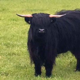

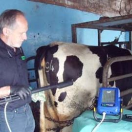
Figure 2. Ovum pickup collection.
Since then, the collection has become very successful, with the use of pioneering ultrasound developments aiding the collection method and, consequently, the oocyte quality harvested. The combination of OPU and IVFEP has developed to such an extent it is commercially available from the main pioneering bovine genetic companies throughout the world.
Advantages of OPU/IVFEP
One of the main benefits for OPU and subsequent IVFEP is successful embryo production, irrespective of the reproductive status of the donor. It can be used in pregnant cows and any that have stopped producing embryos in vivo. It can potentially be used in acyclic cows, and cows with blocked fallopian tubes, genital tract infection or other pathology. Breeders are, therefore, able to produce calves from donors otherwise destined to go barren.
The technique allows the collection of oocytes from cows four to five months into pregnancy. After this stage, the ovary cannot easily be brought in close apposition with the vaginal probe.
Lastly, this technique can speed the production of the next generation of genomically superior offspring as OPU collection can be performed in heifers as young as three to four months of age. Using genomic testing technology to allow identification of superior donors and sires further increases the rate of genetic progress through this embryo production technique.
Additionally, the ability to fertilise with sex-sorted semen allows even greater advantages to be gained. Many of the high scoring genomic bulls are now having their semen sexed, allowing greater choice for fertilisation.

Problems overcome
IVFEP has been used in commercial enterprises, especially in the South American countries, for many years.
Initially, production efficiency was very poor, with small numbers of collected oocytes developing through to the transferable embryo stage. Quality was also an issue, with very low pregnancy rates resulting after transfer.
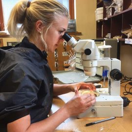
Figure 3. Searching for the cumulus oocyte complex (COC) after ovum pick-up collected.
Further complications associated with IVFEP that occurred once pregnancy was established included increased risk of abortions, large offspring syndrome, an increased rate of hydrops and increased rates of other calf abnormalities. However, these problems and increased risks have largely been overcome with modification of the IVFEP laboratory process. Large offspring syndrome was a problem found to be associated with the use of bovine serum albumin in the culture media. The culture media has been modified to overcome this problem.
he sex ratio of the calves produced have been variable in the past. Some teams had a higher proportion of males than females. Several factors were found to explain the deviation of male:female ratio, including sperm preparation medium and culture medium. Now no difference exists between the numbers of male and female fetuses in most teams and a 1:1 ratio is obtained from quality laboratories.
Process
The two major steps involved in the production of transferable embryos are the on farm/on unit OPU, which is the practical collection of the oocytes, followed by the sterile laboratory IVFEP method that involves laboratory culture of the oocytes, fertilisation of the oocytes and maturation into the bovine embryo.

Cumulus oocyte complex
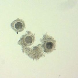
The bovine reproductive egg, the oocyte, is collected in the form of a cumulus oocyte complex (COC). The COC is the oocyte surrounded by the bank of cells (cumulus cells), inside which it sits on the ovary (Figures 3 and 4).
The quality and developmental stage of the COC collected influences the maturation of the oocyte, its fertilisation rate and subsequent development of the embryo, as well as the pregnancy rates of the subsequent transferred embryo.
Figure 4. COC after collection. A good layer of cumulus cells are present.
The COC are classified as follows:
- first grade – more than three layers of compact cumulus cells
- second grade – at least one layer of cumulus cells
- third grade – denuded
- fourth grade – atretic, with dark cumulus cells and signs of cytoplasmic degeneration
Only atretic oocytes are discarded after grading and not started in the maturation process.
Fertilisation
Only a small amount of semen is required to fertilise all the oocytes from one collection. Half a straw is required for one batch of oocytes and they can be divided into batches fertilised by different bulls.

Equipment
The collection equipment consists of an ultrasound machine, a vacuum pump, tubing and a temperature controlled water bath or warming block, plus collection pots
Figure 5
The sensitivity of the ultrasound equipment is extremely important for maximising the number and quality of oocytes recovered. The first studies used a fixed frequency (5MHz) sector probe. Passage to a curve array probe proved more productive (Bols et al, 2004). Probes used today are multifrequency curved linear probes. The multifrequency beam, coupled with its excellent electroncal interpretation, allows visualisation of the small follicle. The image clarity aids aspiration of larger numbers of oocytes for processing.
The ultrasound machine used for OPU is a modified version of one used by vets who perform reproductive examinations during routine fertility visits. The ultrasound probe, which contains the crystals producing the image, is housed in a ridged handle to accommodate a needle guided tubing apparatus. The ultrasound machine, along with its aspiration apparatus and collection tubes and pots, have been perfected over time, allowing collection of good quality COCs
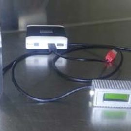

Figure 6
The needle used for aspiration of the COCs has also evolved. The first probes used long 16in specialised needles, which were very expensive and blunted easily. If short needles were used the turbulence caused at the hub applicator junction stripped cells from the cumulus complex rendering a high proportion of the collected COCs unusable. The design has been modified and adapted, and the shorter disposable needles do not cause that problem, are razor sharp and far less expensive.
Suction equipment, such as tubing and fitting, are specially chosen as to be non-toxic to the oocyte and all plastics used are of the very highest quality. Medias have evolved and contain precisely calculated ingredients to feed the oocyte and maintain optimal viability.
Temperatures are crucial, both the ambient temperature of the room and the temperature of the media. The vet undertaking collection can become very warm, as the ambient temperature of the collection area and all equipment is 27°C minimum. The water bath where the COCs are collected into a tube is at 35°C. The laboratory is also kept at these temperatures and a heated stage is used on the microscope.
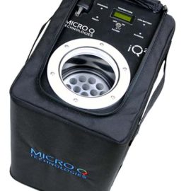
Pots and collection apparatus are sterile, and meticulous procedures are in place to prevent contamination (Figure 6). Often, the processing laboratory is not attached to the collection centre, but can be hundreds of miles away. The COCs are collected into the media. The media containing the collected COCs is then searched under the microscope and the COCs transferred to the maturation media in test tubes. Up to 10 oocytes are placed into each tube and transported via a portable vial incubator
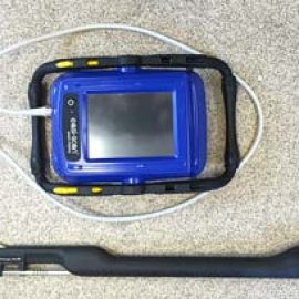

Figure 7
References
- Bols PE, Leroy JL, Van Holder T and Van Soom A (2004). A comparison of a mechanical sector and a linear array transducer for ultrasound-guided transvaginal oocyte retrieval (OPU) in the cow, Theriogenology 62(5): 906-914.
- Brackett BG, Bousquet D, Boice MDL, Donawick WJ, Evans JF and Dressel MA (1982). Normal development following in vitro fertilisation in the cow, Biology of Reproduction 27: 147-158.
- Pieterse MC, Kappen KA, Kruip TA and Taverne MA (1988). Aspiration of bovine oocytes during transvaginal ultrasound scanning of the ovaries, Theriogenology 30(4): 401-413.
- Pontes JHF, Nonato-Junior I, Sanches BV, Ereno-Junior JC, Uvo S, Barreiros RR, Oliveira JA, Hasler JF and Seneda MM (2009). Comparison of embryo yield and pregnancy rate between in vivo and in vitro methods in the same Nelore (Bos indicus) donor cows, Theriogenology 71(4): 690-697.
- Pontes JHF, Silva KCF, Basso AC, Ferreira, CR, Santos GMG, Sanches BV, Porcionato JPF, Vieira PHS, Melo-Sterza FA and Seneda MM (2010). Comparison of oocyte and embryo production among Bos taurus, Bos indicus, and indicus-taurus donor cows, Reproduction Fertility and Development 22(1): 248.
